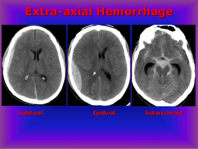
Gambaran CT scan Subdural hematoma, epidural hematoma, sub arachnoid hematoma
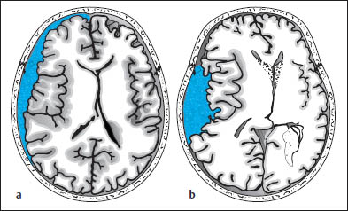
Fig. 1.10 Differentiation between subdural and epidural hematoma. a
Subdural hematoma appears as a hyperdense mass distributed along the
inner table of the skull. It typically has a concave medial border and
extends across cranial sutures. This hematoma overlies the left
frontotemporal convexity and crosses the coronal suture. b
Schematic drawing of epidural hematoma that appears as a more localized,
biconvex hyperdense mass. The dura itself is not depicted.


Komentar
Posting Komentar